About Ultrasound
ImageCare uses state-of-the-art, high resolution ultrasound equipment to evaluate the:
- Heart (echocardiography)
- Abdominal organs
- Pelvic organs
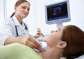
- Thyroid / soft neck tissue
- Scrotum
- Vasculature in the arms and legs
- Extracranial arteries and veins
- Muscles, tendons, ligaments and joints
Musculoskeletal Ultrasound
Ultrasound is increasingly being used as a practical and rapid method of obtaining images of the musculoskeletal system. Musculoskeletal (MSK) Ultrasound imaging uses sound waves to produce pictures of muscles, tendons, ligaments and joints throughout the body. It is used to help diagnose sprains, strains, tears and other soft tissue conditions. Ultrasound is safe and noninvasive. Musculoskeletal ultrasound (MSK) offers substantial benefits to our patients and also to physicians who refer patients to ImageCare for this exam.
- Ideal for evaluation of superficial soft tissue structures, including tendons, ligaments, muscles, cysts, and bursa.
- Accuracy of greater than 90% in multiple series in evaluating full thickness and partial tendon tears, including rotator cuff1-2, Achilles3, peroneals4, and distal biceps tendon5.
- Higher resolution than MRI.
- Allows for dynamic imaging with patient feedback.
- Excellent for evaluation of soft tissues adjacent to hardware that would create artifact with MR and CT imaging techniques.
- More comfortable for patients than MRI – preferred by patients6.
- Less expense than other advanced imaging modalities6.
- Alternative imaging for patients with MR contraindication.
- MR remains standard of care for deep structures, cartilage, and intraosseous abnormalities.
Musculoskeletal ultrasound is available at ImageCare Medical Imaging in Latham, NY. Dr. Michael Cooley of ImageCare specializes in this service. Contact ImageCare Medical Imaging for additional information.
References
- Teefey SA, Hasan SA, Middleton WD, Patel M, Wright RW, Yamaguchi K. Ultrasonography of the rotator cuff: a comparison of ultrasonographic and arthroscopic findings in one hundred consecutive cases. J Bone Joint Surg Am 2000; 82:498–504
- van Holsbeeck MT, Kolowich PA, Eyler WR, et al. US depiction of partial-thickness tear of the rotator cuff. Radiology 1995; 197:443–446
- Hartgerink P, Fessell DP, Jacobson JA, van Hols- beeck MT. Full- versus partial-thickness Achilles tendon tears: sonographic accuracy and charac- terization in 26 cases with surgical correlation. Radiology 2001; 220:406–412
- Grant TH, Kelikian AS, Jereb SE, McCarthy RJ. Ultrasound diagnosis of peroneal tendon tears: a surgical correlation. J Bone Joint Surg Am 2005; 87:1788–1794
- Lobo LG, Fessell DP, Miller BS, Kelly A, Lee JY, Brandon C, Jacobson JA. The role of sonography in differentiating full versus partial distal biceps tendon tears: correlation with surgical findings. AJR Am J Roentgenol. 2013; 200(1):158-62.
- Middleton WD, Payne WT, Teefey SA, Hildebolt CF, Rubin DA, Yamaguchi K. Sonography and MRI of the shoulder: comparison of patient satisfaction. AJR Am J Roentgenol 2004;183:1449–1452
Locations
Select a location:
Technology
At ImageCare, we use a state-of-the-art, high resolution Philips EPIQ and Acuson Sequoia by Siemens. All machines have pulsed wave and color doppler, enabling us to evaluate blood flow within the heart and through the arteries and veins of the arms, legs, neck, and abdomen.
Patient Forms
For your convenience you are now able to view and print pertinent forms online to have ready when you come in for your appointment. The forms below are in .PDF format.
Ultrasound Forms
Patient Preparation
Many ultrasound exams do not require preparation, although you should wear comfortable, loose clothing and may still be asked to change into a gown for some exams. Some exams will require you to fast or to drink water prior to your scan. Our guidelines include:
- Drink 32 ounces of non-carbonated liquids one hour prior to your test.
- Pelvic
- Renal and bladder
- OB’s under 15 weeks
- Nothing to eat or drink after midnight
- Abdominal
- Aorta
- Right Upper Quadrant or RUQ
- Renal Doppler
Frequently Asked Questions
How is the procedure performed?
For most ultrasound exams, you will be lying on your back on an exam table that can be tilted. A clear gel is applied to the area being scanned. This gel is to help the transducer make secure contact with the body and eliminate air pockets between the transducer and the skin. The sonographer then presses the transducer against the skin and moves it back and forth over the area being scanned.
When will my doctor get the results?
After your scan is completed the images will be reviewed by a radiologist who will then interpret and dictate a report. This report will then be sent to your physician within 24-48 hours.